The pituitary gland, often called the “master gland” of the endocrine system, plays a vital role in regulating hormones. However, there are instances where tumours that grow on this gland disrupt hormone balance. This leads to a range of serious health issues, from vision loss and headaches to hormonal imbalances like Cushing’s disease or acromegaly. Traditionally, removing these tumours required a craniotomy, which is a major open-skull surgery. However, a revolutionary technique, the endoscopic endonasal approach, has transformed the surgical landscape. At Snayu Neurosciences, we offer a significantly less invasive and highly effective method for Endoscopic pituitary tumour excision in Kolkata. Therefore, in this article, let us discuss the various benefits of this surgical method.
What is Endoscopic Pituitary Tumour Excision in Kolkata?
Endoscopic pituitary tumour excision in Kolkata is a minimally invasive procedure for removing tumours from the pituitary gland. Using an endoscope, a thin tube with a camera, our surgeons perform the surgery through the nose and sinuses. At Snayu Neurosciences, our neurosurgeon collaborates with our ENT surgeon for the procedure. They rely
They create a small corridor through one nostril to reach the sphenoid sinus, accessing the sella turcica where the pituitary gland sits. Using an endoscope for a magnified view and slender instruments, they carefully remove the tumour. This “keyhole” approach leaves no visible external incision, avoids skull opening, and minimises disruption to brain tissue.
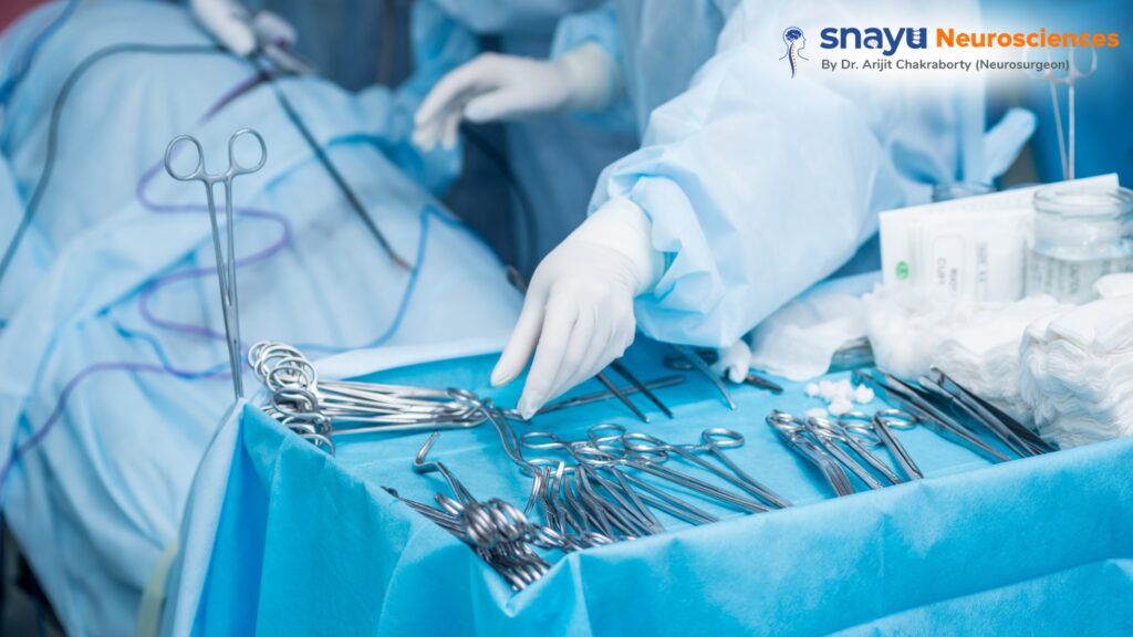
To carry out such an intricate surgery, our surgeons rely on a specific set of tools and technologies to ensure precision and safety. Let us look at the instruments that allow our surgeons to navigate the complex anatomy of the nasal passages and skull base.
Know about the Tools and Their Roles in Endoscopic Pituitary Tumour Excision in Kolkata
| Tool/Technology | Role in Endoscopic Pituitary Tumour Excision |
| Endoscope | Offers a magnified, illuminated view of the pituitary gland through the nostril, enabling the surgeon to see the entire surgical field on a monitor without a large incision. |
| Image-Guided Navigation System (Neuronavigation) | The system acts as a “GPS for the brain,” tracking the surgeon’s instruments in real-time on a 3D model to guide them accurately to the tumour while avoiding vital structures like the optic nerves and carotid arteries. |
| Specialised Microsurgical Instruments | Surgeons use these tools to carefully dissect tumours from surrounding tissue, remove them in small pieces, and repair the site. Their long, thin design enables access to deep surgical areas. |
| Micro-Doppler Probe | This tool is essential for locating critical blood vessels, such as the carotid arteries near the pituitary gland. It helps surgeons confirm their position before dissection, reducing the risk of vascular injury. |
| Intraoperative Imaging (iMRI/iCT) | Enables the surgical team to capture real-time brain images during the procedure, confirming complete tumour removal and accounting for “brain shift,” thus reducing the likelihood of a second operation. |
| Bony Cutting/Drilling Instruments | Used to create a small opening in the sphenoid sinus and sella turcica for tumour access, these tools ensure minimal bone removal and protect delicate structures behind the bone. |
| Hemostatic Materials and Grafts | Grafts fill the space from tumour removal, while hemostatic materials stop bleeding and help repair cerebrospinal fluid (CSF) leaks to prevent infection. |
The Key Advantages of the Endoscopic Pituitary Tumour Excision in Kolkata
The benefits of endoscopic pituitary tumour excision in Kolkata are numerous! Offering a dramatic improvement in patient experience and outcomes compared to traditional open surgery. These advantages can be categorised into several key areas:
- Superior Illumination and Magnification: Unlike a surgical microscope, which offers a limited, fixed view, the endoscope provides a wide-angle, high-definition view with exceptional illumination. The camera is positioned right at the surgical site, allowing for a close-up, highly detailed image of the tumour and surrounding structures.
- Access to Hidden Areas: The endoscope can be manoeuvred into tight spaces and angled to look around corners, giving the surgeon a panoramic view of the sella turcica. This is crucial for identifying and removing every part of the tumour, including extensions that may be difficult to see with a traditional microscope. This “look around the corner” capability helps ensure a more complete tumour removal.
- Preservation of Critical Structures: The magnified view and precise instrumentation allow the surgeon to clearly distinguish the tumour from vital surrounding structures, such as the optic nerves and carotid arteries. This reduces the risk of accidental damage to these critical areas, which could lead to vision loss or stroke.
This Endoscopic Approach Reduces Patient Trauma and Enables Faster Recovery
The minimally invasive nature of the procedure leads to significant benefits for the patient’s recovery. For example, since the surgery is performed through the nose, there are no external incisions, stitches, or visible scars on the face or head. This is a major cosmetic benefit for patients. Furthermore, by avoiding a craniotomy (open skull surgery), the procedure bypasses the need to cut through large sections of skin, muscle, and bone. As a result, patients experience significantly less pain after surgery, reducing the need for strong pain medications.
On the other hand, most patients are discharged within one to two days after the procedure. Especially, compared to a hospital stay of a week or more for a traditional craniotomy. This not only makes the experience less stressful but also reduces healthcare costs. Lastly, with minimal pain and trauma, the recovery is expedited. Patients can often return to work and daily routines within a week or two, a stark contrast to the several months of recovery often required after open-skull surgery.
Lower Risk of Complications in Endoscopic Pituitary Tumour Excision in Kolkata
The inherent nature of the endoscopic approach helps to reduce the risk of common surgical complications. This is possible because at Snayu Neurosciences, our experienced surgeons perform the surgery through a natural orifice. They create a small, clean surgical field significantly reduces the risk of infection. Moreover, this precise, “keyhole” approach and improved visualisation lead to minimal blood loss during the procedure.
Such carefully carried out minimally invasive surgery eliminates the need for blood transfusions. Furthermore, unlike a craniotomy, which may require the brain to be gently moved or “retracted” to access the pituitary gland, the endoscopic approach avoids this entirely. This eliminates the risk of brain swelling, bruising, or other injuries associated with brain retraction.
Snayu Neurosciences Implements a Team-Based Approach for Better Outcomes
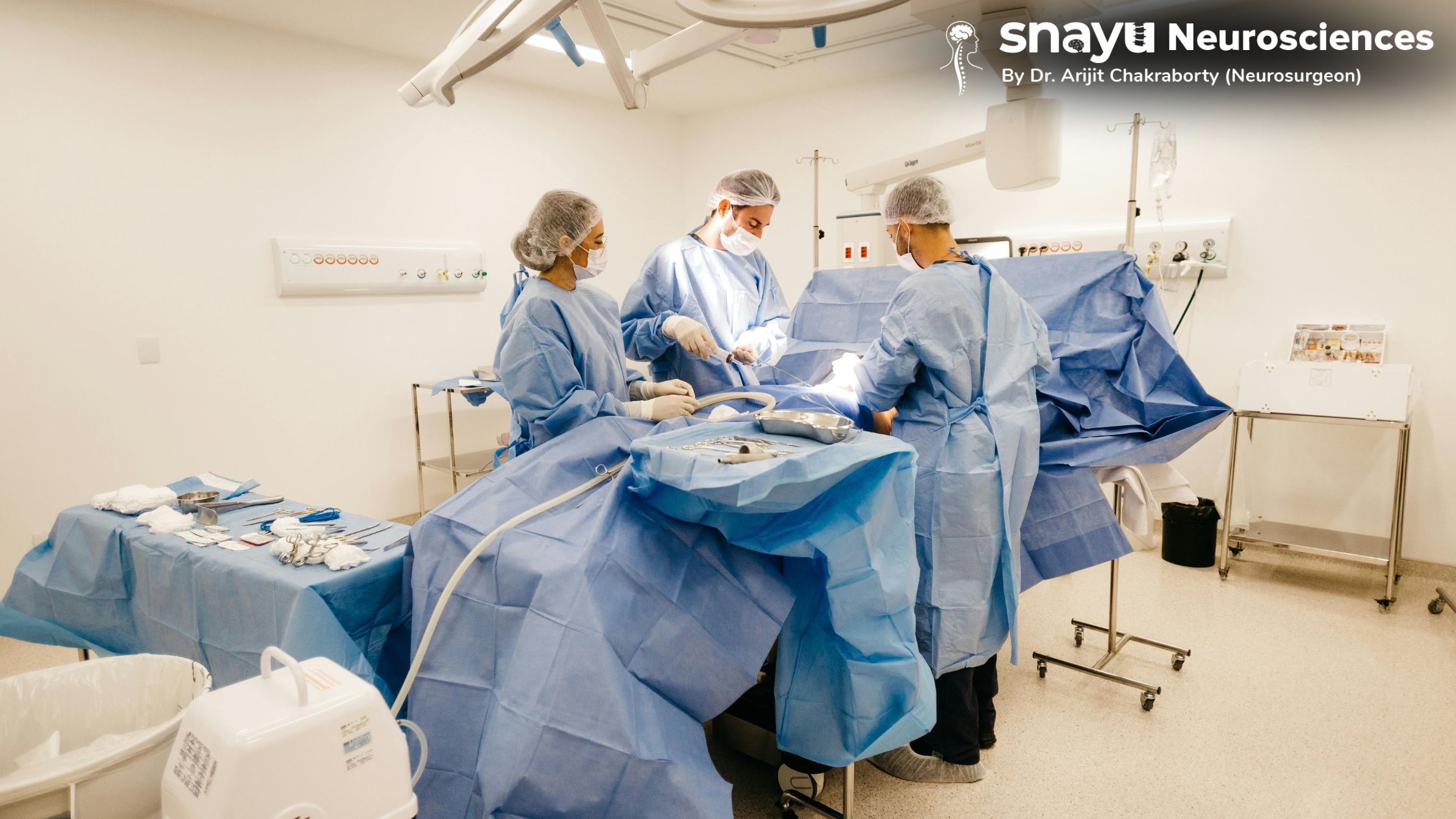
The success of endoscopic pituitary surgery is highly dependent on a collaborative, team-based approach. The procedure is typically performed by our neurosurgeon and an ENT surgeon working together. This combined expertise ensures that the entire procedure. This means from access to tumour removal to reconstruction of the surgical corridor. At Snayu, the whole operation is performed with the highest level of safety and precision.
- The ENT Surgeon’s Role: The ENT surgeon is an expert in nasal and sinus anatomy. They use their knowledge to create a safe, efficient path through the nasal passages and sphenoid sinus to the pituitary gland, ensuring the corridor is wide enough for the neurosurgeon to operate.
- The Neurosurgeon’s Role: Once the path is established, the neurosurgeon takes over. They use their expertise to navigate the delicate structures around the pituitary gland. Meticulously dissecting and removing the tumour while preserving the surrounding nerves and arteries.
Conclusion
Endoscopic pituitary tumour excision in Kolkata represents a major leap forward in neurosurgery. By leveraging advanced endoscopic technology and a team-based approach. We offer a safe, effective, and minimally invasive alternative to traditional open surgery. The ability to achieve a complete tumour removal while significantly reducing patient pain, recovery time. As well as reducing the risk of complications, it has become the gold standard for treating most pituitary tumours. As technology continues to evolve, the “keyhole” approach promises to open up new frontiers in surgical precision and patient care.
FAQs
- Is endoscopic pituitary tumour excision a safe procedure?
- Yes, it is considered the safest and most effective method for removing most pituitary tumours. The procedure’s minimally invasive nature, combined with the use of advanced navigation and visualisation tools, significantly reduces risks compared to traditional open surgery.
- How long will I be in the hospital after the surgery?
- At Snayu, most patients have a very short hospital stay, typically being discharged within one or two days after the procedure. This is a major benefit compared to the longer hospitalisations required for traditional open-skull surgery.
- Will the tumour grow back after it’s removed?
- The goal of the surgery is to remove the entire tumour, but the risk of recurrence depends on the tumour’s type and size. Our neurosurgeon will discuss your specific risk, and you will need to have regular follow-up appointments and imaging to monitor the area for any signs of regrowth.